Existing Patients
(813) 908-8100
New Patients
(813) 474-7201


Digital radiography replaces traditional film with electronic sensors and computer processing to capture dental images. Instead of exposing film packets and chemically developing them, a sensor records the X-ray and converts it into a digital file that appears on a monitor within seconds. This shift from analog to digital changes not only how we see images, but how quickly and accurately clinicians can evaluate oral health.
The core components are straightforward: a low-dose X-ray source, a digital sensor placed near the teeth, and software that receives and displays the image. The software offers tools to adjust contrast, zoom in on areas of interest, and measure dimensions directly on the image. Those capabilities make it easier for clinicians to identify subtle signs of decay, fractures, or bone loss without waiting for film to develop.
At its heart, digital radiography is a technological upgrade with practical benefits. It preserves diagnostic detail while simplifying storage and access. For patients, that means fewer delays during visits and clearer explanations from clinicians when discussing findings and treatment options.
One of the strongest practical advantages of digital radiography is a measurable reduction in radiation exposure. Digital sensors are more sensitive than traditional film, so the same diagnostic-quality image can be obtained using less radiation. This improvement is particularly meaningful for patients who require routine radiographic monitoring over time, including children and those with ongoing dental concerns.
Lower exposure is achieved through both more efficient detectors and software algorithms that enhance image quality. In practice, clinicians can maintain high diagnostic confidence while minimizing cumulative dose. Many professional guidelines endorse the principle of keeping radiation as low as reasonably achievable, and digital radiography is an important tool in meeting that standard.
Beyond dose reduction, the speed of digital imaging eliminates the need to retake images due to development errors. Faster acquisition and immediate feedback help ensure that clinicians capture usable images on the first attempt, further reducing unnecessary exposure.
Digital images offer manipulable detail that film cannot match. Clinicians can adjust brightness and contrast, apply filters, and zoom into specific regions to inspect anatomical structures closely. These on-screen enhancements can reveal early-stage decay, subtle root fractures, and periodontal bone changes that might be harder to discern on standard film X-rays.
Measurements and annotations can be performed directly within the imaging software, which supports treatment planning and record-keeping. For example, practitioners can measure bone height around a tooth or document the exact position of an implant site with greater precision. Those capabilities improve diagnostic consistency across appointments and between team members.
Another diagnostic advantage is digital image standardization. Because files are stored in uniform formats, clinicians can compare images over time side-by-side and apply identical enhancement settings to each image. That makes detecting small changes between visits easier and supports more informed clinical decisions.
Digital radiography integrates seamlessly with modern dental practice management systems, enabling efficient storage and retrieval of images within a patient’s electronic record. Images are filed instantly and indexed to the correct chart, reducing administrative steps and minimizing the risk of lost or misfiled films. That integration speeds up chairside conversations and shortens appointment times.
Because images are digital files, sharing them with specialists, laboratories, or referring offices is fast and secure. Whether a patient needs an oral surgeon consultation or a second opinion, clinicians can send high-quality images electronically rather than mailing physical films or hoping paper copies remain legible. This immediacy improves coordination of care and helps maintain continuity across providers.
For dental teams, the ability to access images concurrently—clinician and assistant, for example—supports collaborative treatment planning. Team members can review the same image, discuss options, and prepare instruments or materials while the patient remains comfortably seated, which makes appointments more efficient and patient-centered.
From a patient standpoint, digital radiography shortens visits and enhances communication. Since images appear on-screen instantly, clinicians can explain findings visually and involve patients directly in the diagnostic process. Seeing a clear image of a cavity or bone change often helps patients understand recommended care more readily than verbal descriptions alone.
Digital systems also reduce the dental office’s environmental footprint. Traditional film processing requires chemical developers and fixers that must be managed and disposed of responsibly. Eliminating those chemicals simplifies clinic operations and avoids potential environmental hazards associated with film processing waste.
Finally, the faster workflow and reduced need for repeats create a smoother, less stressful visit for patients. Taken together—quicker imaging, clearer explanations, and fewer retakes—digital radiography contributes to a more positive and efficient dental experience.
Adopting digital radiography is part of providing modern, evidence-based dental care. For residents of Tampa and surrounding communities, having access to up-to-date imaging means earlier detection of problems and more precise treatment planning when issues arise. Clinicians who embrace digital tools can offer care that is both patient-focused and technically robust.
In our practice, we use digital radiography to support thorough examinations and clear communication. High-quality images help us identify concerns early and document progress over time. When appropriate, we combine digital images with clinical examination and other diagnostics to form a complete picture of oral health and develop tailored care plans.
Arevalo Dental Studio balances technological capability with an emphasis on patient comfort and understanding. By integrating digital imaging into routine care, we aim to make visits faster, safer, and more transparent—helping patients make informed decisions about their oral health.
Digital radiography is an important advancement in dental imaging: it reduces exposure, enhances diagnostic clarity, streamlines record-keeping, and improves the patient experience while eliminating film-processing waste. These benefits collectively support better clinical decisions and more efficient, patient-centered care.
If you have questions about how digital radiography is used in your care or would like to learn how modern imaging might affect your next visit, please contact us for more information. Our team is happy to explain the process and what you can expect during an imaging appointment.
Digital radiography uses electronic sensors and computer processing to capture dental images instead of film and chemical development. A sensor placed near the teeth records X-rays and converts them into a digital file that appears on a monitor within seconds, eliminating the wait and handling required for film. This change improves image availability and facilitates faster clinical decision making.
The core system includes a low-dose X-ray source, an intraoral or extraoral sensor, and software that displays and stores images. Software tools allow clinicians to adjust contrast, zoom, and measure structures directly on the image, which enhances visualization compared with static film. Digital files also simplify storage and retrieval, making long-term comparisons and record keeping more efficient.
Digital sensors are more sensitive than traditional film, so diagnostic-quality images can usually be obtained with less radiation, which is particularly important for children who may need periodic monitoring. Clinicians follow the ALARA principle (as low as reasonably achievable) to minimize exposure by using the lowest effective settings, appropriate shielding, and careful positioning. Pediatric protocols are adjusted to the child’s size and clinical needs to limit dose while maintaining diagnostic value.
For pregnant patients, routine radiographs are often deferred when possible, but necessary imaging can be performed with precautions such as abdominal shielding and strict adherence to dose-limiting techniques. Decisions about imaging during pregnancy are made collaboratively between the patient and clinician, weighing the diagnostic benefit against any potential risk. If you are pregnant or think you might be, let the clinical team know so they can tailor the approach accordingly.
Digital images can be manipulated on-screen to reveal details that might be difficult to see on film, such as early decay, subtle root fractures, or changes in bone height. Clinicians can adjust brightness and contrast, apply filters, and zoom into regions of interest to enhance diagnostic clarity, which supports earlier detection and more precise assessments. Built-in measurement and annotation tools help quantify findings and improve communication among providers.
Standardized digital files also enable side-by-side comparisons of images taken at different times with identical enhancement settings, making it easier to detect small changes over sequential visits. This consistency supports more reliable monitoring of conditions like periodontal bone loss or lesion progression. The combination of enhanced visualization and standardized records leads to more informed treatment planning and follow-up.
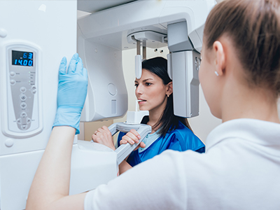
A typical visit for digital radiography is quick and straightforward: the clinician or assistant positions a small sensor inside or near your mouth while a handheld or wall-mounted X-ray source briefly emits a pulse to capture the image. Each exposure lasts only a fraction of a second, and the resulting image appears on a monitor almost immediately for review. The team will ask you to remain still and may provide a lead apron for added protection.
If an image is unclear, the immediate feedback from the monitor allows the clinician to retake the exposure right away, reducing the chances of missed information at a later date. After acquisition, the clinician will review the images with you, explain findings using on-screen tools, and discuss recommended next steps. This chairside review helps patients understand their oral health and the rationale for any proposed care.
Digital sensors are more efficient at converting X-ray photons into image data than traditional film, so lower radiation levels are typically sufficient to produce clear diagnostic images. Software algorithms further enhance image quality, allowing diagnostically useful detail to be visible even at reduced exposure settings. Immediate feedback and fewer retakes also lower cumulative dose by ensuring usable images are captured on the first attempt.
In addition to sensor sensitivity and software, clinicians use technique improvements—such as proper collimation, exposure settings based on patient size, and positioning aids—to limit exposure. Professional guidelines endorse these measures to keep radiation as low as reasonably achievable. When combined, these practices maintain diagnostic confidence while minimizing patient dose.
Yes. Digital radiographs are stored as electronic files that can be securely exported and shared with specialists, laboratories, or referring dentists, enabling faster consultations and coordinated care. Common file formats and standards such as DICOM facilitate compatibility across imaging systems, so receiving providers can view high-quality images without loss of detail. Electronic transfer is faster and more reliable than mailing physical films.
When a referral or second opinion is needed, the practice can transmit images via secure, HIPAA-compliant channels or provide patients with a copy on request. This ability to quickly share images helps streamline treatment planning and reduces delays associated with traditional film transfer. Patients should always be informed about how their records will be shared and may request copies for their personal records.
Digital radiographs are integrated into the patient’s electronic record and stored on secure servers with routine backups to protect against data loss. Practices typically use encrypted storage and access controls so only authorized team members can view or modify images, and audit logs track access for accountability. Regular software updates and IT safeguards are important to maintain security and compliance with privacy regulations.
Redundancy and off-site backups help ensure images remain available for future comparisons and treatment planning, while retention policies follow professional and legal guidelines. Patients can ask the practice about specific data protection measures if they have concerns. Clear communication about record-keeping and privacy builds trust and helps patients understand how their information is secured.
Digital radiography itself does not automatically change the recommended frequency of X-rays; imaging intervals are determined by individual risk factors, clinical signs, and professional guidelines. Factors such as age, history of decay, gum disease, and symptoms guide the clinician’s recommendation for bitewing or full-mouth series timing. The advantage of digital systems is that they make comparisons over time simpler, which can refine decisions about monitoring intervals.
In routine care, clinicians perform a risk assessment at each exam and use that information to tailor imaging schedules to the patient’s needs. For patients with stable, low-risk oral health, radiographs may be needed less frequently, while those with higher risk or active problems may be monitored more often. Discussing your individual risk profile with the clinical team will clarify the appropriate imaging plan for you.
Digital radiography shortens appointments by producing images instantly and reducing the need for repeats, which makes visits more efficient and less stressful. On-screen images support clearer communication because clinicians can show and annotate findings directly, helping patients visualize issues such as cavities or bone changes. The immediate availability of images also speeds up treatment planning and referral coordination when needed.
Removing film processing eliminates chemical waste and simplifies clinic workflows, which has positive environmental implications and reduces handling delays. For many patients, the combination of faster imaging, clearer explanations, and fewer retakes creates a more comfortable, transparent experience. These benefits contribute to better-informed decisions and smoother appointments.
Arevalo Dental Studio adopts digital radiography to combine diagnostic precision with patient-centered care, using technology that enhances visualization while minimizing exposure. Digital imaging supports thorough examinations, consistent monitoring over time, and clearer chairside communication so patients can better understand findings and treatment options. The practice integrates these images into secure electronic records to streamline workflows and collaboration with other providers when necessary.
For patients in Tampa and surrounding communities, access to modern imaging means earlier detection of problems and more precise treatment planning when issues arise. Clinicians and team members use the tools to document progress and tailor care plans to each patient’s needs, balancing technological capability with comfort and clear explanation. If you have questions about how imaging will be used during your visit, the team can explain the process and the safeguards in place.